
Centers of Excellence
All radiodiagnostic examinations & mammography with modern machines, specialized radiologists, responsibility and safety.
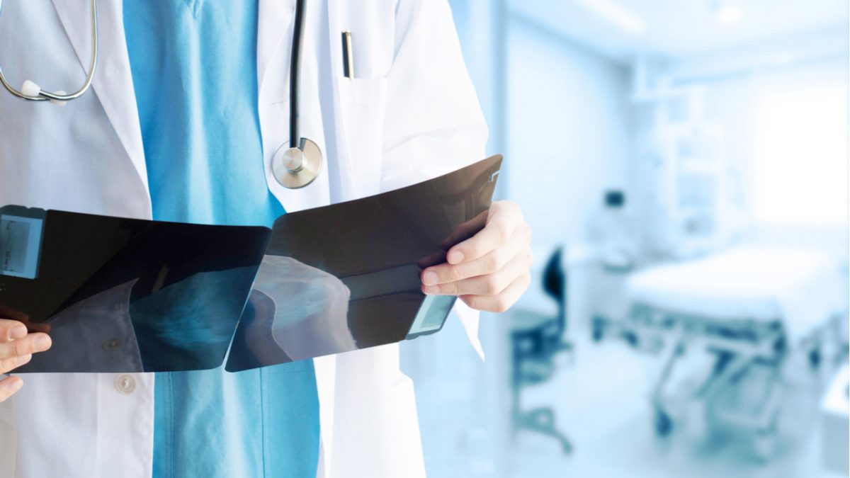
The Diagnostic Radiology Departments perform the entire range of conventional radiology examinations and mammographies. Special imaging sectors (interventional radiology, interventional neuroradiology, pediatric radiology, cardiac hemodynamic imaging and cardiophysiology imaging) operate under the care of the Department of Diagnostic radiology for the proper function of all devices. These special procedures are performed under the scientific management of specialized stuff.
MITERA Hospital Diagnostic Radiology Department is equipped with:
Digital Procedures
All devices, including the CR units, are connected to the HIS and RIS digital file systems of both hospitals. Furthermore, all radiography exams are performed based on a work list, which limits to a minimum any errors when recording patient details.
All digital radiography images are stored in the PACS and are available to physicians at all times for reading them and making comparative assessments, thus limiting the need for films. Doctors reports are also stored into each patient’s medical record.
Outpatients receive printouts of their images and reports, as well as a CD/DVD with the digital images of their exams. All digital images are stored in the Department’s digital archive.
Digital imaging advantages:
• It practically eliminates technical errors associated with X-ray views and the need for frequent re-exposures to ionizing radiation.
• It minimizes the radiation dose.
• It ensures secure storage of diagnostic images.
• It offers fast and convenient access to diagnostic data for referring/attending physicians, even remotely using safe internet connection.
The following examinations are performed:
• All X-rays of the body (chest, abdomen, head, limbs and spine).
• All specialized tests for the digestive and genitourinary systems using contrast mediums. Specifically, these include assessment of the esophagus, stomach, duodenum, small intestine (per os and enteroclysis) and large intestine, assessment of stomies and bariatric procedures, assessment of the biliary tract, intravenous urography and retrograde pyelography and urethrocystography, fistulography and hysterosalpingography.
• Cephalometric and panoramic dental and jaw X-rays
Moreover, the cutting-edge technology of the flat-panel digital fluoroscopy device at HYGEIA Hospital assists in performing full-body spine radiographies for assessment of scoliosis and full-length radiographies of limbs for assessment of leg length discrepancies. The same machine is also used for rotational radiographies and cone-beam tomography for 3D reconstructions.
Centers of Excellence

Centers of Excellence

Centers of Excellence

Centers of Excellence