
Centers of Excellence
We perform all imaging tests that relate to the prevention and diagnosis of breast conditions.
It is staffed with fully trained and qualified healthcare professionals who use the latest technology to provide immediate and accurate results to patients and physicians, either as part of a check-up or for further investigation of findings arising from a physical examination of the breast.
All digital images (mammographies and ultrasounds) are stored in the PACS and are available to physicians at all times for evaluating them and making comparative assessments. Diagnosis reports are also stored electronically into each patient’s medical record.
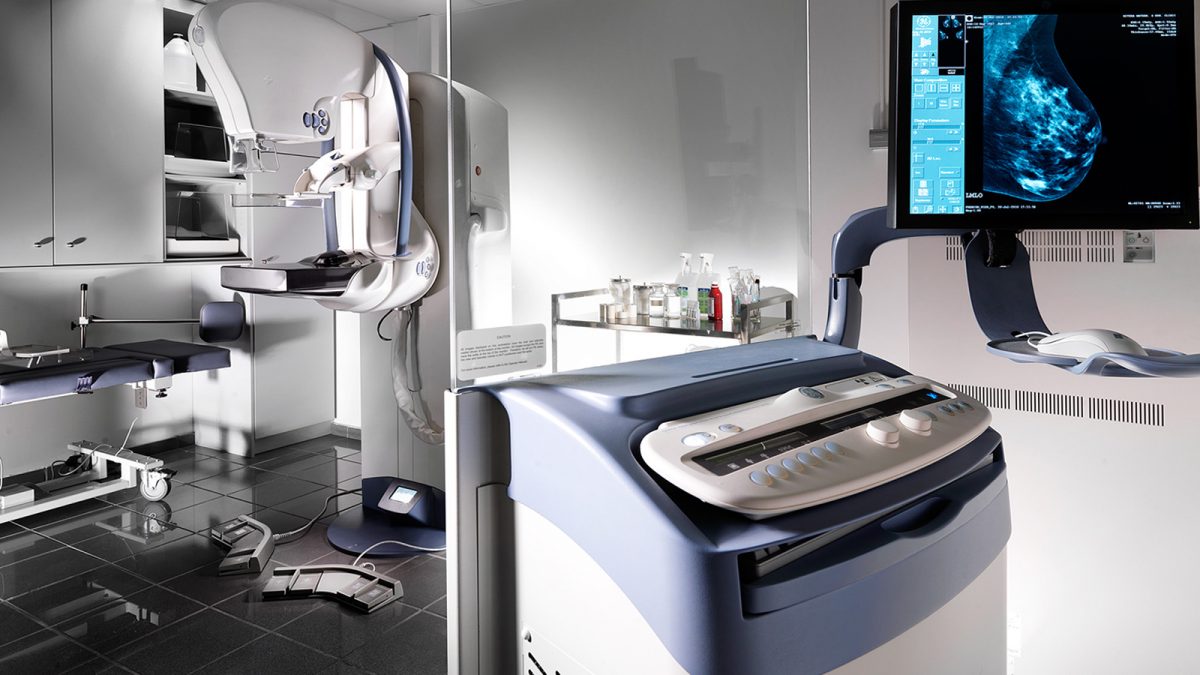
Mammography
The Center is equipped with two latest-generation Senographe Essential GE digital mammography units, with a built-in flat-panel sensor. The two digital mammography units have been set to automatically determine the appropriate amount of emitted kV and mAs, based on the density and size of the breasts, as well as the pressure exercised on them
The radiologists present during the exam assess the mammograms immediately. If they deem it necessary to further investigate an area of interest or a specific mammography finding, they notify the technicians accordingly. The technicians take the images that have been selected from a series of body position views, either as spot or magnified compression views (using special compressors for such images), with the aim of assessing and evaluating the findings.
Once a medical opinion has been formed, the radiologists inform the patients in person on the test results and provide the necessary clarifications and recommendations for possible further clinical and imaging assessment, while they also answer any questions.
Ultrasound/Elastography
The Center is equipped with a digital Siemens Acuson S2000 high-definition ultrasound system, used exclusively for breast examinations. The ultrasound system is exclusively and constantly available to a qualified radiologist for further investigation of mammography or clinical findings, with the aim of providing fast and accurate imaging results.
The device is equipped with elastography technology, which provides information on the elasticity of breast ultrasound findings, contributing to their optimized imaging representation.
Stereotactic biopsy
Vacuum-assisted stereotactic breast biopsies may also be performed at the Breast Center. It is a minimally invasive procedure performed with local anesthesia under mammography guidance. Small tissue parts that present mammographic interest are extracted and aspired using 7 or 10g needles, depending on the case. It is usually performed to remove suspect calcification clusters.
The tissue is then sent to pathology for analysis and the diagnosis is as accurate as with a biopsy performed via surgical excision, but without the accompanying adverse effects of a surgical procedure.
Mammographic or ultrasound-guided placement of surgical guidewire: If the surgical excision of non-palpable lesions or calcifications is deemed necessary, surgeons are guided with the use of guidewires, which are placed under mammographic guidance. Specialized radiologists perform multiple guidewire placements at the Breast Center safely and accurately, working closely with the experienced and specialized technical staff.
If the suspect finding is detected during an ultrasound, then the surgical guidewire is placed by the qualified radiologists under ultrasound guidance.
Breast puncture-core biopsy under ultrasound guidance: It is a well known fact that an ultrasound is the screening of choice for the visual representation of palpable findings. If a histological identification of said findings is deemed necessary, then there is the option of a breast puncture using fine need aspiration (FNA) or core biopsy, i.e. extraction of part of the suspect tissue, using a larger needle (14g). The Breast Center is equipped with a latest generation Siemens Acuson S2000 ultrasound system for exclusive use. The system offers the possibility of immediate histological examination of suspect findings, under ultrasound guidance by qualified radiologists, using any of the methods deemed necessary.
INSTRUCTIONS
According to the updated recommendations issued by the American Cancer Society (ACS) in 2015, the guidelines for breast cancer screening for women at average risk are as follows:
– Regular screening mammography must start at 45 years of age
– Women aged 45 to 54 years should be screened annually*
– Women 55 years and older should transition to biennial screening for life*
– Routine clinical breast examination or self-examination is not considered necessary
Note that the American College of Radiology (ACR) and the Society of Breast Imaging (SBI) have not accepted the new ACS recommendations and continue to propose annual breast screening for life, starting at the age of 40.
* Women should have the opportunity to begin annual screening at the age of 40 and continue with annual screenings for life, after consulting their physician.
![]()
Breast Scan: Pan-European AI platform for advanced breast cancer screening.
Centers of Excellence

Centers of Excellence

Centers of Excellence

Centers of Excellence