
Centers of Excellence
Apart from all types of diagnostic studies, the whole range of percutaneous (diagnostic and therapeutic) procedures are performed, on any part of the body.
The unified HYGEIA-MITERA CT & MRI Department is equipped with the latest technology, offering advanced medical services 24/7/365. All types of imaging studies can be performed at the CT and MRI Department, after contacting the Department Reception by phone or in person.
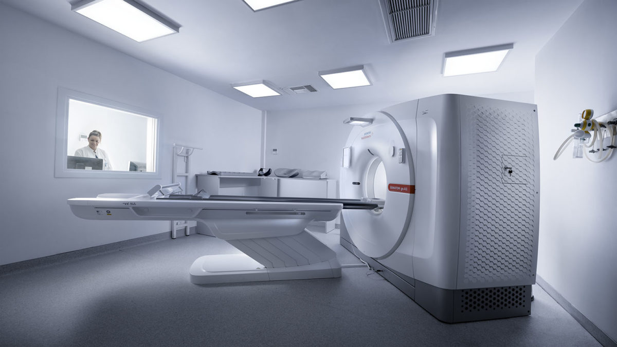
Four (4) multi-slice CT scanners are installed in the Department. Among them, one 64-slice dual-source scanner (SIEMENS DEFINITION) at HYGEIA Hospital. The latter is ideal for performing special and complex scans, such as CT coronary angiography (non-invasive angiography), CT angiography of the entire body and CT colonography (virtual colonoscopy). A 64-slice CT scanner is also installed at MITERA Hospital. The Department implements protocols for reducing patient radiation exposure during a CT scan by up to 40%.
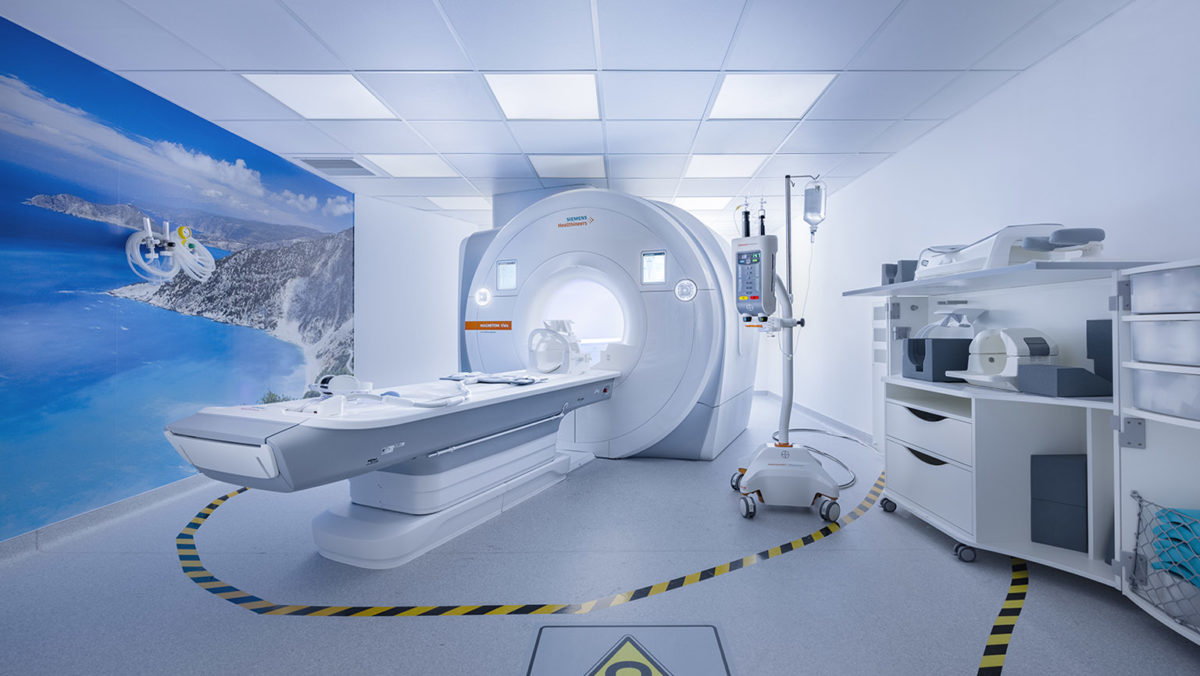
Four (4) advanced technology MRI scanners are installed in the Department. Specifically, two 1.5 Tesla MRI scanners as well as a state-of-the-art 3 Tesla MRI scanner are installed at HYGEIA Hospital. The latter is ideal for imaging of the musculoskeletal and nervous systems. The cutting-edge technology of the 3 Tesla system assists in performing advanced imaging studies, such as multiparametric prostate MRI, MR spectroscopy of the brain, functional MRI (fMRI) of the brain and MR Tractography.
The Department is a center of excellence in MRI of the Breast in Greece. MRI-guided interventional breast procedures are also performed. A 1-Tesla open MRI scanner is also installed at MITERA Hospital, which is the most powerful open-type MRI device in Greece.
This CT study examines the human body blood vessels, such as the brain arteries, as well as the thoracic and abdominal aorta and their branches. The examination is performed similarly to any other CT scan. However, the intravenous administration of iodinated contrast medium is always necessary.
The examination is safe, painless, bloodless and lasts just a few seconds (10-20 sec, depending on the test). The images are sent to special workstations, where the radiologist uses dedicated software to reconstruct and study them.
Over the last few years, CT angiography has replaced conventional angiography for investigating a great variety of conditions of the vessels, such as aneurysms and stenoses
It is a non-invasive, painless CT method used to evaluate the trachea and bronchial tree. The examination lasts a few seconds and does not require any preparation on the part of the patient. Thin CT slices are obtained, which are then processed by the radiologists at special workstations. Then 2D and 3D reconstructions are created to provide images of the tracheobronchial tree and show any potential pathological findings in it.
It is an advanced CT study used to detect cancer and polyps in the large bowel. It is a non-invasive, safe and painless method, which does not require hospitalization or administration of anesthesia. At the same time, it also allows screening of the rest of the abdominal organs during the same examination. A very low radiation dose is used.
As opposed to conventional colonoscopy (whereby the colonoscope must be inserted through the entire large bowel), in this examination a small catheter measuring approximately 10 cm is inserted in the rectum, through which gas (carbon dioxide) is introduced to gradually expand the colonic lumen. The duration of the study is no longer than 15-20 minutes. To ensure a successful examination and optimal evaluation, adequate preparation (colon cleansing) is required, similar to that for a conventional colonoscopy. The test is suitable for patients who must undergo colon cancer screening, but are unable to do so or do not wish to have a colonoscopy, as well as in the cases when conventional colonoscopy was not completed. The detection rates for clinically significant polyps are comparable to those of conventional colonoscopy.
CT Coronary Angiography is the most effective non-invasive diagnostic imaging test for assessing the coronary arteries that supply blood to the heart. It is a fast, safe and painless method which does not require hospitalization.
A CT Coronary Angiography could prove useful for:
The examination may also prove useful for people with two or more risk factors associated with the development of coronary disease, such as high blood cholesterol levels, obesity, arterial hypertension, smoking, family history of coronary disease and diabetes mellitus. In these cases, the test may lead to fast diagnosis or exclusion of coronary disease. It must be performed with the consent of the attending physician, the cardiologist or the internist.
The examination does not require any special preparation. However, patients must follow some special instructions before the test. The duration of the whole procedure is no longer than 15-20 minutes, while the actual test (heart scan) does not exceed 20 seconds. The examination is performed in the dual-source multi-slice CT scanner. Its advanced technical features assist in performing reliable studies, even on patients with tachycardia or a history of atrial fibrillation.
It has been widely accepted that CT Coronary Angiography has a high negative prognostic value, i.e. it can rule out coronary disease with an accuracy of almost 100%. Furthermore, its diagnostic accuracy is similar to that of conventional coronary angiography.
This examination has been developed over the last few years with the aim of assessing the small bowel. It is mainly used to diagnose and evaluate the extent of idiopathic inflammatory bowel disease and, especially, Crohn’s disease. In the last years, it has replaced in most cases the conventional enetroclysis performed through fluoroscopy. Other indications for a CT Enterography/Enteroclysis study include: investigation of blood loss, with possible origin at small bowel, as well as detection and evaluation of small bower tumors.
The actual study (image scan) does not last longer than 20 seconds, i.e. as long as an abdominal CT.
What you need to know about this examination:
• Enterography: You will be asked to receive a specific quantity of a special solution orally prior to the exam, in order to expand the small bowel lumen. Therefore, you would better arrive at the Department one hour prior to your scheduled exam.
• Enteroclysis: during this study, a solution is administered directly into your small bowel via a nasal catheter. The catheter is previously placed under fluoroscopic guidance. Therefore, you have to arrive about half an hour prior to the scheduled time of the exam, for the catheter placement.
For both studies:
• No preparation is required apart from a 6-hour fast.
• Because the solution has a cathartic action, it is recommended to postpone any activities outside the house during the rest of the day after the exam and to assure easy access to a bathroom.
• A contrast medium will be administered to you intravenously during the exam.
A shift in the size of a lung nodule is a significant criterion for evaluating its biological behavior (malignant or benign). Monitoring a lung nodule with 3D volumetric measurement is considered more accurate and reliable compared to measuring only one or two of its dimensions. The CT-MRI Department of HYGEIA Hospital, is equipped with state-of-the art software for automatic volumetric measurement of pulmonary nodules, allowing reliable detection of significant changes in the nodule and early diagnosis of a malignant lesion.
Segmental volumetric measurement provides useful information to surgeons prior to performing a partial hepatectomy, since it accurately calculates the amount of liver parenchyma that will be preserved after the scheduled surgical procedure. The The CT-MRI Department of HYGEIA Hospital is equipped with advanced software for localizing and measuring the volume of the focal lesions and liver lobes. Intravenous administration of iodinated contrast medium is required before undergoing the test.
CT Urography is an optimal technique for evaluating the urinary system, highly-sensitive in detecting stones, congenital disorders and kidney masses. It is, also, indicated for investigating hematuria, offering useful information to the clinician.Intravenous administration of iodinated contrast medium is required before undergoing the test.
Before scheduling any examination, please note:
• A CT scan is an examination based on the implementation of X-rays. Therefore, in case of known or possible pregnancy, please notify the Reception or the Department staff.
• You are advised to arrive at the Department 20 minutes before the time of your scheduled examination, to allow for all administrative matters.
• If you have been scheduled for a lower abdominal CT scan, you will be asked to arrive one hour before the time of the actual exam. The reason for that is to have time for the administration of an oral solution, enabling better contrast enhancement of the bowel during the exam.
• You must bring with you any previous test results (CT or MRI) for comparison. If the previous exams had been performed at our Department (from 2009 onwards), then you do not need to bring them with you, as they will be stored in the Hospital’s digital archive.
• For some tests (e.g. those performed as part of oncology patient monitoring), a special medication (iodinated contrast medium) must be administered intravenously for contrast enhancement of the internal organs and vessels.
• If you have a history of allergic reaction to previous administration of iodinated contrast medium (e.g. during a CT or intravenous pyelogram) or a history of allergies in general, you must notify the Department Reception while scheduling your appointment for the examination. Anti-allergic preparation instructions may be given to you if deemed necessary.
• If you are over 70 years old, you will be asked to submit recent (within the last two months) blood urea and creatinine test results. The same applies if you are a diabetic or experience any problems with your renal function.
• If you are taking the drug metformin (marketed under various trade names) for your diabetes, please notify the staff of the department when you schedule your appointment or just before you undergo your CT examination. Besides, you will be asked so during your visit, just before undergoing the examination.
• In all other events, you may take any medications prescribed to you by your physician.
• If you have any further questions, you may contact the Department Reception.
Magnetic Resonance Imaging (MRI) is based on the magnetic properties of protons in matter, when these undergo the effect of strong magnetic fields. Before undergoing an MRI scan, it is absolutely necessary to rule out that the patient has specific metallic materials or medical devices (such as pacemakers) implanted, since these would not make it safe for them to be placed in the strong magnetic field created by the device.
Individuals with claustrophobia, who cannot remain still or cooperate during the MRI scan, may have the exam performed under mild sedation or at MITERA Hospital, which is equipped with an open MRI scanner. The best option is proposed by the Department radiologists, after consulting the patient’s physician and discussing it with the patient. Mild sedation may be administered by an anesthesiologist, after suitable planning.
The intravenous administration of a special (paramagnetic) contrast medium is required for certain MRI scans. This is generally safe and any adverse effects from its administration are extremely rare. However, special precautions are required in the case of pregnant women, breastfeeding mothers or patients with severe kidney failure
All conventional and specialized MRI exams are performed at HYGEIA and MITERA hospitals, in the most suitable scanner for each case.
• Brain
• Pituitary gland
• Petrous bones
• Οrbit
• Paranasal sinuses
• Head
• Neck
• Chest
• Cervical spine
• Thoracic spine
• Lumbar spine
• Sacral spine
• Upper abdomen
• Retroperitoneal space
• Lower abdomen (pelvis)
• Magnetic resonance cholangiopancreatography (MRCP)
• MRI of bones and soft tissue
• MRI of joints (temporomandibular, shoulder, elbow, wrist, hand, hip, knee, ankle & foot)
MRI is an established imaging technique to investigate all the human body organs and systems for a wide range of conditions. It contributes greatly in the evaluation of certain conditions affecting the nervous and musculoskeletal systems, while it keeps gaining ground in newer applications, such as assessment of blood vessels, heart and other anatomical structures. MRI offers detailed anatomical information on normal and pathological tissues, but it may also provide functional information. This information is quite significant and often decisive for proper diagnosis, which leads to the choice of the most suitable treatment.
The precise mapping of tissues is of crucial importance in the preoperative plan for patients, as well as in their reassessment to evaluate the treatment results. Due to the absence of ionizing radiation, MRI is suitable for young patients, especially when monitoring requires regular reexaminations. MRI can be safely used on patients for whom a CT scan with IV administration of contrast medium is unsafe due to allergic reaction to iodine or due to renal failure. The contrast mediums used in MRI are not nephrotoxic (apart from gadolinium), while allergic reactions are extremely rare and generally mild.
This examination assists in evaluating the blood vessels of the brain, spinal cord, neck and other regions of the body.
It is a painless and non-invasive method that provides images of the blood vessels on any part of the human body. In the majority of cases, it permits to avoid the conventional angiography, which is relatively painful and not free of complications. It is used to diagnose stenoses, obstructions, aneurysms or other vascular conditions with great accuracy.
This exam requires intravenous injection of a contrast medium (gadolinium), apart from a brain MRA which usually does not require administration of contrast medium.
Magnetic resonance cholangiopancreatography is a MR application that provides 3D images of the biliary system and pancreatic duct in detail. It is a painless, non-invasive method that does not use ionizing radiation and does not require the administration of contrast medium. It can be used to detect stones in the biliary tree, as well as neoplasms or other rare conditions.
Modern MRI scanners have the capability of performing accurate evaluation of the iron deposition in organs such as the liver and myocardium, offering useful information to clinicians, especially in the case of multitransfused patients. MRI is currently considered a reliable, safe and non-invasive method for evaluating iron deposition. As proven by various studies, the results it offers are linearly correlated with those of direct evaluation of liver iron load through biopsy.
Modern MRI scanners offer the option of not only detecting, but also quantifying liver fat deposition. Many studies conclude that there is a linear correlation between the quantity of fat measured by an MRI scanner and that estimated from liver biopsies. This method may prove useful in cases of diffuse hepatic steatosis, as well as other liver diseases related to alcohol or drug abuse.
It is a modern MR application that offers useful information about the urinary system. Since is does not use ionizing radiation, it is quite suitable for children and younger patients in general.
These are advanced applications in the field of neuroimaging that are performed in modern generation MR scanners. Perfusion MR assesses the blood supply of the brain at a microvascular level. This method assists in investigating hyperacute (very recent) ischemic brain conditions and neoplasms, as well as the advanced differential diagnosis of neoplasms and other pathological conditions.
It is an advanced imaging technique that provides information on a molecular level. It can contribute to a more accurate identification of brain lesions, such as tumors.
It is an advanced imaging technique that provides information regarding the functional activity of the brain. It offers the possibility of evaluating the function of vital and significant brain centers, such as speech and movement (e.g. movement of the hand, foot, etc). It has many applications, but the most common one is preoperative assessment of patients with brain tumors. In this case, the exam may offer neurosurgeons information regarding the anatomical relationship between the tumor and certain vital brain centers.
It is an advanced imaging technique that depicts the neural tracts of the brain (e.g. corticospinal tract, visual tract, etc). The method is applied in cases of malignant brain tumors, motor neuron diseases and other pathological conditions.
The HYGEIA and MITERA Imaging Department is equipped with advanced technology and can perform all types of Cardiac MRI scans. It is also staffed with qualified personnel, which have contributed to the clinical application of Cardiac MR imaging internationally and have received recognition through a series of scientific articles, lectures and distinctions.
An MRI scan is a non-invasive diagnostic technique. The patient is not exposed to any radiation (X-rays or gamma rays).
Nowadays, magnetic resonance offers the possibility of evaluating a wide range of heart conditions such as:
• Assessment of the anatomy of the heart and the major thoracic blood vessels. Some of the most common clinical applications include assessment of the aorta for dissection and aneurysms, assessment of possible pericardial thickening and constrictive pericarditis, as well as evaluation of cardiac and pericardial tumors.
• Various types of cardiomyopathies, such as acute, dilated, hypertrophic and infiltrative cardiomyopathy.
• Assessment of the left and right ventricle function of the heart.
• Quantitative assessment of the blood flow, which can be used to perform quantitative measurements of cardiac supply and valve dysfunctions, even on patients with left to right communication.
Detection of myocardial viability. In patients with a history of ischemia or myocardial infarction, MRI using a contrast medium (gadolinium) can accurately depict the extent of the infarction, even if it only relates to subendocardial layers.
Assessment of myocardial perfusion in stress (vasodilation) and rest for evaluation of induced ischemia and coronary disease.
Imaging of the origin and proximal course of the coronary arteries to rule out disorders in the origin of the vessels and/or significant proximal coronary disease.
It is a highly sensitive method for diagnosing breast cancer, without the use of ionizing radiation. It is usually performed as a supplemental imaging study following mammography, clinical exam or ultrasound. It is a specialized imaging exam, which must be performed when there are appropriate indications. It is very effective in diagnosing many breast conditions, while it may provide answers to a wide range of clinical problems. It lasts approximately 20 minutes. Women of reproductive age are advised to undergo the exam between the 7th and the 14th day of their menstrual cycle, so as to limit the possibility of false-positive results. For the same reason, possible hormonal replacement therapy (HRT) in post-menopausal women is better to be interrupted 6-8 weeks before the examination.
HYGEIA Hospital’s MRI Department is a center of excellence in Magnetic Resonance Imaging and the only center in Greece that can perform MRI-guided interventional breast procedures.
These are special MR techniques for evaluating the small bowel and its conditions. They are quite useful in diagnosing and evaluating the extent of idiopathic inflammatory bowel disease and, especially, Crohn’s disease. Having the added benefit of not using potentially harmful radiation, these examinations are recommended for the regular monitoring of patients with inflammatory disease, especially of a younger age. They are also useful for diagnosing small bowel tumors. Each study has a duration of approximately 30-40 minutes. MR Enterography requires the oral administration of approximately 1.5 liters of a special solution, during one hour prior to the examination. In the case of MR Enteroclysis, the solution is administered directly to the small bowel via a special nasal catheter introduced under fluoroscopic guidance. The most suitable study (enterography or enteroclysis) is selected after discussion between the the Department doctors and the patient’s attending physician
MR Arthrography is a special magnetic imaging technique that is performed with the administration of paramagnetic contrast medium (gadolinium) into the joint via a fine needle. This method is very effective for diagnosing disorders in intra-articular structures (such cartilage and ligaments).
In the CT and MRI Department may also be performed diagnostic biopsies and aspirations on any part of the body. With this method, either a needle is inserted in the area of the lesion with millimeter precision and fluid is collected for cytology testing (Fine Needle Aspiration – FNA) or a small part of tissue is collected and examined by pathologists (Core Needle Biopsy – CNB).
Department’s medical staff, also performs percutaneous drainage of abscess and other fluid collections using catheters, as well as nephrostomy. Furthermore, CT-guided ablation of bone and other tumors may be performed. Biopsies have to be scheduled in advance and instructions must be obtained.
Multiparametric MRI of the prostate gland combines the anatomic and functional techniques of magnetic resonance, significantly improving the accuracy of diagnosing prostate cancer.
Specifically, it provides useful information on the existence of multiple cancer foci in the prostate gland, enables the performance of targeted transrectal biopsy, the exact staging of the disease, the optimal treatment planning (surgery, radiotherapy), the evaluation of the treatment result and the detection of local relapse. It is a significant tool for optimal patient management. The examination has duration of approximately 40 minutes and the Department is equipped with a latest generation 3 Tesla MRI scanner that uses a surface (not endorectal) coil.
MRI is a highly sensitive method for diagnosing breast cancer. In many cases, suspicious breast lesions are only visible in an MRI and cannot be detected in conventional tests (mammography or ultrasound). In these cases, the Department physicians can perform MRI-guided localization or biopsy of these breast lesions so as to enable the identification of their exact nature.
Centers of Excellence

Centers of Excellence

Centers of Excellence

Centers of Excellence