
Centers of Excellence
High quality diagnostic examinations in newborns and children up to 16 years of age.
The entire range of pediatric diagnostic examinations is offered by the MITERA Children’s Hospital Pediatric Imaging Department which covers the imaging needs of all pediatric patients, from neonates to 16-year-olds. The Department also performs Fetal MRIs.
The staff consists of 6 specialized radiologists, 14 radiographers, 2 nurses and secretaries.
The diagnostic exams include ultrasounds, conventional radiographs, fluoroscopic studies (micturating cysteourographies, upper GI studies, enemas, etc), fluoroscopic video swallowing studies, CT scans and MRIs. Apart from the outpatients, the Department also covers the imaging needs of inpatients (neonates, infants and pediatric patients) of MITERA Hospital’s clinics and ICUs.
Multiple examinations are performed in the Pediatric Imaging Department, with aim to reach the accurate diagnosis in various medical conditions which affect neonates, infants and Children:
These examinations use small radiation doses to depict human body structures, such as the lungs and the bones. They are also used to examine the intestines or detect a foreign object, often swallowed by young Children.
During these diagnostic examinations, a contrast medium (such as barium or iodinated contrast medium, which are both visible on plain films) is administered to the child, either orally or through a tube. This achieves contrast enhancement of anatomical structures, such as the gastrointestinal tract or the urinary system. A video swallowing study which analyzes the swallowing process in children with swallowing difficulties may also be performed.
This is an examination that uses ultrasounds to depict parts of a child’s body, without any biological burden. Certain areas can be examined only up to a certain age. For example, a brain U/S may only be performed while the anterior fontanelle (the part of the cranium over the temple that is soft when palpated) is patent, and a hip U/S until the hip joint is ossified. The pediatric U/S secretaries will address any questions you may have about the examination and they will advise you regarding the preparation required.
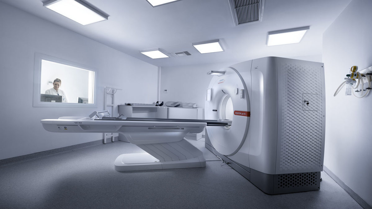
A computer system takes more complicated images of internal body structures and is used when there are indications requiring more detailed assessment of these structures. In children, the decision to proceed with a CT scan is not taken lightly. There have to be specific clinical evidence to this direction, since the method carries greater radiation burden. In every case, special low-radiation dose pediatric protocols are applied for children.
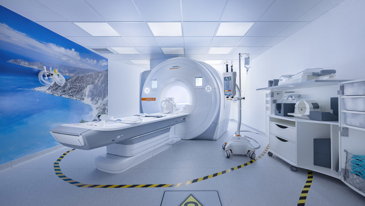
The scanner uses a magnetic field, and not ionizing radiation, to depict the various systems in children, without any biological burden. The exam lasts longer than a CT scan, so mild anesthesia (sedation) is applied to younger or uncooperative children in order to keep them still.
Fetal MRI is a modern imaging technique which complements fetal ultrasound.
Where is it performed? At MITERA Hospital’s MRI Department using a high-field open-type MRI scanner (1,0 – Tesla).
Preparation: The expectant mother should fast for 4 hours prior to the exam and must bring with her the fetal ultrasound – scan. Are the baby and the mother safe? It is universally accepted that Fetal MRI is entirely safe, especially when performed after the first trimester.
Procedure: The mother lies down comfortably during the exam, while she has the option to be accompanied in the MRI suite during the scan. All Department staff are working hard to provide these circumstances that will make mother feel calm and relaxed.
Duration: The duration of the exam depends on the cooperativeness of the fetus. When the fetus is calm, the exam lasts 30 minutes on average.
Benefits: The Fetal MRI focuses on confirming sonography findings but most importantly on detecting other sonographically occult anomalies. It is not affected by the fetal projection or the maternal body habitus, while it assesses structural development according to gestational age.
Which pregnancies should undergo fetal MRI?:
1. Those with abnormal findings in the fetal brain, chest, abdomen or spine in the fetal ultrasound
2. Complicated monochorionic twin pregnancies
3. When there is the family history of central nervous system anomalies in other children or pregnancies
4. Fetus with congenital intrauterine infection.
When should a Fetal MRI be performed?
It is usually performed during the 23rd week of gestation. When follow up is required this should be done on the 32nd week of gestation.
This is an examination that studies the anatomy and function of the kidneys and the drainage system, and it is usually performed under anesthesia. The child receives intravenous hydration prior to the exam.
It is a special evaluation of the digestive tract and specifically the small intestine, which is performed after administration of a special solution. The child must drink a mannitol solution, which looks and tastes like water, at a steady pace, one hour before the examination. For this reason, this examination cannot be performed under sedation. The child must follow a liquid diet on the previous day.
It is a special assessment of the anatomy and function of the heart using magnetic field.
Radioisotope tests are performed to study anatomical body structures and check their function, e.g. urinary system.
• The equipment includes state-of-the-art devices, adjusted to the imaging needs of young patients, both in terms of aesthetics and technology. The protocols applied are based on the international pediatric imaging guidelines. All contemporary pediatric imaging guidelines are followed, through continuous education and updates; the key aim is reliable diagnosis, with as low as possible biological and psychological burden on the young patients. Low-dose protocols designed for children and based on the international radiation protection guidelines for pediatric patients (ALARA, Image Gently) are in place for all examinations requiring ionizing radiation (conventional & specialized radiographs and CT scans).
• Furthermore, the picture archiving and communication system (PACS) was launched in 2009 and was integrated in all Departments in 2011, so that all test results are since digitally archived, with the option of retrieval and digital transfer.
• Conventional Pediatric Radiology Department: It is equipped with a Siemens YS10 digital X-ray machine, a Siemens ICONOS R-200 digital fluoroscopy machine and two portable Siemens POLYMOBIL PLUS SN 21345 and SN 21344 X-ray machines.
• Pediatric Ultrasound Department: It is equipped with a GE LS6 unit and two portable F2-GE units.
• CT Department: It is equipped with a latest-generation Siemens Sensation 64-slice CT scanner.
• MRI Department: It is equipped with a high-field (1.0T) open-type Philips Panorama MRI scanner, which offers high-quality images in an Ambient Experience environment. Prior to the examination, children may play with a toy simulator, to better understand and accept the examination procedure, performing themselves examinations on special dolls and watching a relative story unfold on TV.
• The open MRI scanner offers children the possibility of visual and physical contact with their parents/guardians during the examination. Moreover, the Ambient Experience environment creates a friendly and relaxed atmosphere during the examination, projecting cartoons and playing music.
• It is also possible to have pediatric tests performed at HYGEIA Hospital, i.e CT scans, MRI scans and PET-CT scans and nuclear studies.
A CT scan uses ionizing radiation and depicts various body parts of the child with the use of a computer. The CT scanner is a device in the shape of a ring, with a large hole in the center, through which a bed moves in and out. With the modern multi-slice CT scanners, each examination lasts from a few seconds to a few minutes, so anesthesia is usually avoided. Special attention is given to reduce the radiation burden on children. Low-dose CT protocols are set, the area to be scanned is limited, and the examination is performed only when it is absolutely necessary, e.g. on the chest to check the lungs and on the bones in the event of injury.
Why does your child need a CT scan?
Your child may need to undergo a CT scan so that an area of the body may be studied in detail and a treatment method may be suggested.
What does the exam include?
• You are welcome to stay in the CT room with your child during the exam, but you must wear a special lead apron to protect yourself from radiation. The apron will be given to you by the radiographer who will perform the scan. If there is suspicion of pregnancy, you must inform the CT staff prior to the examination and you will not be allowed to enter the room during the scan. The same question will be posed to girls of reproductive age for legal purposes, without having the intention to place the patient or her parents in an uncomfortable situation.
• The child will lie on the CT scanner bed and will be placed in the correct position by the radiographer in charge, who will then leave the room to enter the CT control room. As part of the examination, the scanner bed will move to reach the middle of the CT scanner. During the examination, which usually lasts for a few minutes, the scanner makes a noise in order to produce the images. The radiographers can communicate with you and your child via intercom. The pediatric radiologists often attend the examination. As soon as it is over, the radiographer will pull out the scanner bed and the child may get up.
Are there any risks?
• CT scans are performed on children only when the benefits outrange the potential risks of radiation. In children, the radiation dose is set to the minimum required to ensure diagnostic image quality.
• Your child must remain still during the examination. Mild anesthesia (sedation) is recommended for uncooperative or young children to ensure that they will not move. The anesthesia is administered via a small catheter which is placed in a vein on the child’s hand, under the supervision of a pediatric anesthetist.
• In some CT scans, it is also necessary to administer a contrast medium through a vein, in order to study certain areas more accurately. This medication is not recommended for children with renal insufficiency or children who are allergic to iodine.
Preparing for the exam
• If necessary, you will receive special instructions from the Department secretaries when confirming the appointment for the examination. If your child will undergo anesthesia, you will receive special instructions from the pediatric anesthetist the day prior to your appointment. You must follow the instructions closely to ensure that the examination will be performed on scheduled time.
• Sleeping on the previous night
It is advisable to cut down on your child’s sleeping hours prior to the examination. For example, put them to sleep 2 hours later than their usual time and wake them up 1 hour earlier.
• Fasting
All children who undergo mild anesthesia must have consumed their last meal or beverage (milk) at least 4 hours prior to the examination and water up to 2 hours before, always according to the pediatric anesthetist’s instructions. Children must not consume anything the final 2 hours before the examination. If these instructions are not followed, the examination will have to be postponed.
• You will be given a consent form prior to the examination, whereby you will agree and give permission for the examination to be performed. For younger children, there is a CT toy simulator available in the Department. This helps children to understand how the images are taken and what the doctors are looking for, while they may also perform their own scans on special dolls.
What happens after the examination?
• If the examination was performed without anesthesia, you may depart immediately and receive the results on the following working day. If the examination was performed with sedation, you will wait in a room with your child for 20 to 30 minutes, until he recovers completely and the pediatric anesthetist gives permission for you to leave.
Children who have undergone sedation may experience nausea for the next 24 hours following the examination. In this case, limit your child’s food and liquid intake until he feels better. It is also possible for the child to feel fatigue after sedation and for the next 24 hours, so it is best to limit activities that require alertness and coordination, such as cycling. However, do not feel concerned if you child seems drowsy or irritable after sedation. Simply monitor them until they completely recover.
• Do not hesitate to contact the CT-MRI Department if you have any questions or concerns.
• The MRI scanner uses a magnetic field and not ionizing radiation to produce images of a child’s body. It is the most suitable imaging technique for the brain, spine, abdomen and joints.
• The MITERA MRI Department has set up a pleasant and friendly environment for young patients and their guardians. For children of a borderline age (4-7 years) there is an MRI toy simulator available, so that anesthesia may be avoided. Children get to perform MRI scans on special dolls, while they also watch a story on a screen about how each doll stood still and took great images.
• The child, along with his guardian, is then escorted to the MRI room, where a relaxing environment, the Ambient Experience, projects cartoons with soft lighting and music. Children may also bring over their own CDs to listen to the music of their choice during the examination.
• The MITERA Hospital MRI scanner is a high-field (1-Tesla) open magnet. Children lie on it without having to enter inside the conventional tube. They are in visual contact with their environment and can also be in physical contact with the parent or the guardian accompanying them into the room. Like all MRI scanners, a loud repetitive tapping noise can be heard during the exam.
• Children lie on the scanner and a special headset is placed on their ears to better listen to the music and phase out the noise of the scanner. Special headsets, compatible with the MRI scanner, are also placed on infants and very young children who undergo sedation, for protection against the noise. The exam usually lasts 30 to 40 minutes.
• You will be given a consent form to fill out and sign prior to the examination, whereby you will agree and give permission for the examination to be performed, as well as a consent form for the administration of sedation. If you have any questions while filling out the forms, you can discuss them with the Department nurses or the pediatric anesthetist.
Note that prior to entering the MRI room, you will be asked whether you or your child carry metal implants or a pacemaker that are incompatible with the magnetic field.
Why does your child need an MRI scan?
The exam may be deemed necessary by clinical physicians, so that they may obtain detailed information on the part of the body being examined and plan treatment accordingly.
What is the procedure for an MRI?
• As soon as you arrive at the Hospital, proceed to the MRI Department secretariat (level 2). You are welcome to stay in the MRI room during the examination, provided it is not performed under anesthesia. However, if the guardian is in the first trimester of pregnancy or has metal implants or a pacemaker, they must notify the Department staff. Do not hesitate to bring your child’s favorite CD, to listen to it during the examination.
• The child will need to lie on the MRI bed and a coil (a component that looks like a ring) will be placed around the area to be examined. In a Brain/Head MRI, a coil resembling a helmet is placed around the head, without touching it. When the child is in the right position, the technologist performing the examination will move the bed to the middle of the scanner and will then proceed to an adjacent area, the control room. However, they keep having visual and audio contact with the child and the examination room. As soon as the examination starts, a loud repetitive noise resembling drums will be heard. Both the child and the guardian will wear headsets to phase out the noise and listen to the music better.
• The radiographer will be in contact with the child via a microphone and the child will have an emergency button at hand to press in case of emergency. When the examination is over, the technologist will pull the bed outwards and the child will be able to get up.
What happens after the examination?
If the examination was performed without anesthesia, you will be able to depart immediately. The pediatric radiologists will then study the images and the official report will be ready on the following working day; the secretariat will inform you about the exact time slot.
Are there any risks?
• There are no risks associated with the MRI scans. Children do not experience any pain or inconvenience whatsoever during the examination.
• MRI scans cannot be performed on individuals with metal implants or pacemakers, as they use a very powerful magnetic field. For children wearing braces, parents must consult the orthodontist to verify whether the materials are compatible with the magnetic field. Otherwise, the braces should be removed. During the examination, children must not wear clothes with zips or metal buttons. If so, they will need to be removed; a hospital gown will be provided.
• Prior to the examination, the MRI nurse will hand to the guardian the consent form and ask for possible metal implants in the child’s body or possible allergies. You will be asked to sign a consent form that includes all these details. You will also be asked to remove any metal objects, such as jewelry, watches, coins, keys, and also credit cards and mobile phones, prior to the examination. You may store these items in special lockers in the preparation room.
• The child must remain completely still throughout the examination, so mild anesthesia (sedation) may be required if the child in uncooperative. In some MRI scans, the use of an intravenous contrast medium in required for contrast enhancement of the area being examined. A small catheter will be placed on a peripheral vein, usually in the hand, prior to the infusion.
• Rest assured that we have collectively taken all the necessary steps so that the procedure is as pleasant and effective as possible for both you and your child. However, some examinations may last longer than anticipated, so please bear in mind that delays may occur.
• Sedation is recommended for infants or younger children who are uncooperative and cannot stay still during the examination. This is performed by pediatric anesthetists. Preparation instructions are given so that the child does not consume any food, water or other fluids a few hours prior to the examination . In the event of a CT scan, which lasts only a few minutes, the physician discusses the possibility of performing the examination without sedation, provided the child can remain still and cooperate to a certain extent. For infants undergoing a CT scan, the examination may be attempted as soon as the child falls asleep, usually after feeding.
Prior to the examination
The Hospital’s nurses place a venous catheter on a peripheral vein, usually in the upper limb, which is used to administer intravenously the sedation drugs.
• Sleeping on the previous night
It is advisable to cut down on your child’s sleeping hours prior to the examination, but it is best for the parents to decide this, since they are aware of their child’s reaction to insomnia.
• On the day of the examination
Wake up your child an hour earlier than the usual. Also try to keep them awake during the drive to the hospital.
• Fasting (solids and liquids)
Do not breastfeed your baby 2 hours prior to the appointment for the examination. Babies and children on milk (formula or cow milk) must not have consumed any solids or liquids 4-5 hours prior to the appointment. Clear liquids, such as water, may be consumed up to 2 hours prior to the examination.
After the examination
You must remain in the Department until your child is fully awake (usually 20 to 30 minutes after the end of the examination). The pediatric anesthetists will give you instructions for the following few hours. In case of an emergency, do not hesitate to contact the MITERA Children’s Hospital Emergency Department.
Examinations under anesthesia
For pediatric imaging tests requiring anesthesia, the Hospital’s pediatric anesthesiologists are available 24/7, both for emergencies and scheduled appointments. Anesthesia applied is usually sedation, most commonly for children younger than 4 or 5 years of age. The anesthesia is performed and monitored by pediatric anesthesiologists, who are present throughout the examination. They use a special monitor (INVINO PRECESS) to observe the child’s vital signs until he has fully recovered.
In most cases, hospitalization is not required when a child undergoes an examination under anesthesia and he may leave the Hospital 30 to 45 minutes
Centers of Excellence

Centers of Excellence

Centers of Excellence

Centers of Excellence