
Centers of Excellence
Αρχική σελίδα MITERA Hospital Medical Departments & Clinics Ocular Oncology Center Pediatric and Adult
Leading Unit in Europe
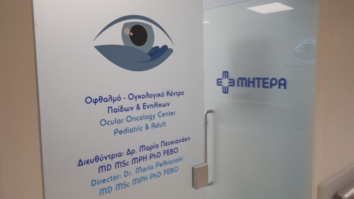
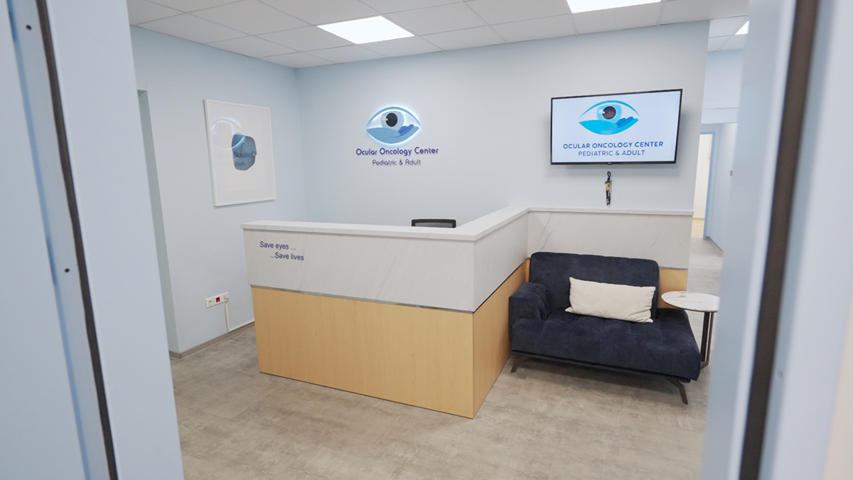
The Ocular Oncology Center for Children & Adults of MITERA, a member of the Hellenic Healthcare Group (HHG), specializes in the diagnosis and treatment of ocular tumors in children and adults. We apply the latest diagnostic and therapeutic methods, providing individualized care to each patient.
We combine leading medical expertise with advanced technologies.
OPTOS Silverstone, the most powerful tool yet for examining the retina, is the only ultra-widefield imaging device with integrated swept-source...
OPTOS Silverstone, the most powerful tool yet for examining the retina, is the only ultra-widefield imaging device with integrated swept-source OCT.
OPTOS Silverstone produces a 200° single-capture retinal image of unrivaled clarity in less than ½ second and enables optomap-guided OCT scanning across the retina and into the far periphery.
Routine optomap use enhances pathology detection, disease management, and clinic flow.
Now, with Silverstone, examination of the retina from vitreous through the choroidal-scleral interface is faster and easier than ever.
OPTOS Silverstone SS OCT, the most powerful tool for examining the retina in patients with ocular Malignancies.
The Ocular Oncology Centre of MITERA Hospital is The only Ophthalmic center in Greece with this Equipment
See more
OPTOS Silverstone offers the following benefits:
Image Views
optomap Image Modalities
OPTOS Silverstone Fluorescein Angiography ultra-widefield, ultra-high resolution angiography, enables a simultaneous pole-to-periphery view of the retina of the eye. This supports practitioners...
OPTOS Silverstone Fluorescein Angiography ultra-widefield, ultra-high resolution angiography, enables a simultaneous pole-to-periphery view of the retina of the eye. This supports practitioners in diagnosing ocular pathology that may first present in the periphery of the retina, resulting in better patient outcomes.
The Ocular Oncology Centre of MITERA Hospital is The only Ophthalmic center in Greece with this Equipment
This allows the entire retinal vasculature to be imaged during the dye transit by a non-contact method
OPTOS Silverstone Indocyanine Green Angiography (ICG) ultra-widefield, ultra-high resolution angiography, enables a simultaneous pole-to-periphery view of the retina of the eye. This...
OPTOS Silverstone Indocyanine Green Angiography (ICG) ultra-widefield, ultra-high resolution angiography, enables a simultaneous pole-to-periphery view of the retina of the eye. This allows the entire retinal vasculature to be imaged during the dye transit by a non-contact method.
It can yield additional information helpful in differentiating amongst choroidal tumors allowing more accurate treatment planning and follow-up.
The Ocular Oncology Centre of MITERA Hospital is the only Ophthalmic center in Greece with this Equipment
This supports practitioners in diagnosing ocular pathology that may first present in the periphery of the retina, resulting in better patient outcomes.
RetCam Envision sets the standard in pediatric ocular imaging, delivering brilliant images and very important diagnostic test in children with...
RetCam Envision sets the standard in pediatric ocular imaging, delivering brilliant images and very important diagnostic test in children with retinoblastoma. Absolutely necessary for ocular oncology specialty. RetCam makes pediatric ocular imaging easy, and sets a new standard in the field.
Enhanced illumination with an ultra-wide field of view makes RetCam Envision the leader in image quality.
RetCam Envision’s fluorescein angiography (FA) option is a diagnostic aid to examine the circulation of the retinal blood vessels and assist with the detection of anatomical abnormalities
The Ocular Oncology Centre of MITERA Hospital is The only Ophthalmic center in Greece with this Equipment
With Light Shaping Technology patent users experience a greater visualization with brilliant color images. These unique features result in images that provide a higher level of confidence for patient evaluations and consultations. Brilliant color images can be viewed in full-screen mode for maximum visualization of detail, enabling earlier detection of eye diseases and timely intervention.
The RetCam Envision’s lens portfolio, detachable portrait lens and detachable 130ο lens, provides the best partner for anterior and posterior imaging. Together with the Light Shaping Technology, lenses give focus across the entire 130º field of view, improved peripheral illumination, and better illumination of darkly pigmented retinas. Furthermore, lenses can be removed easily for cleaning and disinfecting with minimal downtime. It provides the ability to visualize the absence or presence of blood flow within the eye.
RetCam Envision’s fluorescein angiography option is a diagnostic aid to examine the circulation of the retinal blood vessels and assist with the detection...
RetCam Envision’s fluorescein angiography option is a diagnostic aid to examine the circulation of the retinal blood vessels and assist with the detection of anatomical abnormalities. An essential test in retinoblastoma patients defining the extent of the disease and distinguishing this tumour from other pediatric ocular condition.
The Ocular Oncology Centre of MITERA Hospital is The only Ophthalmic center in Greece with this Equipment
World-wide famous, Swiss-made Haag-Streit Slit Lamp meets professional photography in the unique combination of BX-900. Making use of the excellent...
World-wide famous, Swiss-made Haag-Streit Slit Lamp meets professional photography in the unique combination of BX-900.
Making use of the excellent microscope optics and a DSLR camera, the user has the power to capture astonishing details of the Anterior Chamber and the Cornea pathologies and drastically contribute with diagnosing and treating them.
The Haag-Streit BX-900 is a no-compromise slit lamp-imaging instrument designed an important diagnostic test in patients with suspicious lesions on the anterior segment of the eye.
The Ocular Oncology Centre of MITERA Hospital is the only Ophthalmic center in Greece with this Equipment
The BX 900 slit lamp is a no-compromise slit lamp imaging instrument designed to assist ophthalmic healthcare providers.i
Benefit from the best camera systems on the market. The Haag-Streit BX 900 slit lamp can be equipped with various professional camera systems thanks to its universal adapter, which is elegantly located above the slit lamp microscope.n
Envisu, is a hand-held OCT solution with an intended use to aid in the diagnosis of physiological and pathologic conditions...
Envisu, is a hand-held OCT solution with an intended use to aid in the diagnosis of physiological and pathologic conditions of the eye. It has an essential role in retinoblastoma management, particularly in tracking submillimeter tumors. It is vital in retinoblastoma care to confirm retinoblastoma recurrence, influencing the decision to provide further treatment, and establioshing the tumor response to therapy.
Quickly capture and observe brilliant, high-resolution, and high-density optical coherence tomography (OCT) images where you need it.
The gentle, non-contact Envisu C2300 system has a significant impact on pediatric ocular oncology, especially on retinoblastoma cases.
Permits precise tumor monitor for growth and detection of subclinical or invisible retinoblastoma.
The Ocular Oncology Centre of MITERA Hospital is the only Ophthalmic center in Greece and whole Europe with this Equipment
Spectralis is a world-renowned device for fundus tomography. Heidelberg Spectralis Optical Coherence Tomography, an expandable diagnostic imaging platform wich combines...
Spectralis is a world-renowned device for fundus tomography.
Heidelberg Spectralis Optical Coherence Tomography, an expandable diagnostic imaging platform wich combines scanning laser fundus imaging with high-resolution OCT imaging, and the enhanced depth imaging OCT (EDI-OCT), are helpful in the diagnosis, treatment planning and monitoring response of ocular and periocular tumours in adults and children.
It is designed to provide high quality pictures of the retina macula, the optic nerve and other optical tissues and help to diagnose Diabetic Retinopathy, Age-related Macular Degeneration and several other pathologies.
Multi-spectral fundus imaging (Multicolour Module Imaging) on Heidelberg Engineering OCT Spectralis, an imaging acquired with three simultaneous laser beams at...
Multi-spectral fundus imaging (Multicolour Module Imaging) on Heidelberg Engineering OCT Spectralis, an imaging acquired with three simultaneous laser beams at different depths of the ocular fundus, a novel technique that can be used to highlight different layers within the retina simultaneously, provides three monochromatic images that can reveal topographic changes not visible with conventional color fundus photography, crucial diagnostic tool help find tumour boundaries and the extent of macular involvement, which may be useful for assessing visual impact.
Fundus imaging modalities, including high-resolution images and video sequences that show vessel filling, flow and leakage and are essential tools...
Fundus imaging modalities, including high-resolution images and video sequences that show vessel filling, flow and leakage and are essential tools for the investigation of tumour circulation.
PERMITS IMAGING EXCELLENCE The entire eye is visible with an exceptional level of detail, a crucial diagnostic element for the...
PERMITS IMAGING EXCELLENCE
The entire eye is visible with an exceptional level of detail, a crucial diagnostic element for the diagnosis of eye tumors.
ABSolu combines the best image quality, the highest measurement accuracy in its category, with excellent ergonomics.
The ultrasound is the primary diagnostic test that confirms the diagnosis of choroidal melanoma as well as the size, thickness (apical height), basal dimension, extraocular extension (such as scleral nodules), and document growth during the follow-up of a suspicious nevus/small melanoma.
Ultrasonography in retinoblastoma patients is very helpful in accessing the tumor and in differentiating retinoblastoma from other conditions
Innovative B-scan probe 20MHz, with five concentric rings that direct the ultrasound according to the requirements of the examination and offer 70% greater examination depth and increase the detail of the imaging by 25%.
A-scan probe, standardized by Ossoinig at 8MHz Specialized A-scan probe, standardized by Ossoinig at 8MHz, ideal for clinical examination of...
A-scan probe, standardized by Ossoinig at 8MHz
Specialized A-scan probe, standardized by Ossoinig at 8MHz, ideal for clinical examination of ultrasound absorption characteristics of tumors, carcinomas, melanomas and other pathological findings.
Absolu Standardized probe, important tool on diagnosis of eye tumour
ABSolu combines the best image quality, the highest measurement accuracy in its category, with excellent ergonomics.
UBM 50 MHz ultrasonic biomicroscopy probe, ideal for the detailed examination of the anterior segment of the eye (cornea, iris,...
UBM 50 MHz ultrasonic biomicroscopy probe, ideal for the detailed examination of the anterior segment of the eye (cornea, iris, circular body) with unsurpassed resolution and tools for quantifying various sizes in the image (angle, angular opening, lens vault, etc.).
ABSOLU Ultrasound biomicroscopy (UBM) 50MHz linear, an important tool for assessing and the management of anterior segment tumours. Selected cases of advanced retinoblastoma, UBM appears to represent a valuable tool in the precise evaluation of anterior extension of the disease.
Improved UBM technology makes it possible to diagnose structures behind the iris that other technologies cannot visualize. The new signal processing mode offers increased resolution and invasiveness.
ABSolu combines the best image quality, the highest measurement accuracy in its category, with excellent ergonomics.
Humphrey Field Analyzer 3 combines everything that you can imagine making it simpler, faster and more reliable than ever before...
Humphrey Field Analyzer 3 combines everything that you can imagine making it simpler, faster and more reliable than ever before in a Humphrey
Humphrey Visual Fields, used to detect blind spots or scotomas in the visual field. The nature of such blind spots (shape, size, depth) can point towards different disorders of the eye, optic nerve or visual pathway in the brain, including also tumours.
Quicker, simpler, more dependable than ever Liquid Lens technologies: One lens, less time The automatic Liquid Trial Lens reduces patient setup time by automatically loading each patient correction from the previous examination.
The improvements in HFA3 from a Humphrey add to the standard that is reliable that thousands of clinics already depend on for critical diagnoses. The new model still provides when and where you require it.
STATPAC™ statistical software provides comprehensive analysis by comparing results to proprietary age-normative and glaucoma databases, enabling effective monitoring of visual field changes over time.
SOLIX is a new technology built upon a proven foundation of high-speed Spectral Domain OCT. This FullRange platform empowers practitioners...
SOLIX is a new technology built upon a proven foundation of high-speed Spectral Domain OCT. This FullRange platform empowers practitioners to identify and manage numerous pathologies from the front of the eye to the back for a vastly superior diagnostic experience.
Visionix Optical coherence tomography angiography (OCTA) is a noninvasive imaging modality used to diagnose and manage retinal and choroidal vascular disease.OCTA has the ability to detect subtle lesions that are not identifiable during clinical examinations. OCTA has a role in understanding tumour vascularity.
SOLIX delivers multiple tools for a new generation of disease management that improve throughput and enable superior patient care:
SOLIX is a new technology built upon a proven foundation of high-speed Spectral Domain OCT. This FullRange platform empowers practitioners...
SOLIX is a new technology built upon a proven foundation of high-speed Spectral Domain OCT. This FullRange platform empowers practitioners to identify and manage numerous pathologies from the front of the eye to the back for a vastly superior diagnostic experience.
OCT provides detailed images of ocular tissue structure, enabling early detection of anomalies and monitoring of ocular disease progression.
AS-OCT has proved to be an indispensable diagnostic tool in diagnosing and managing conjunctival and corneal pathologies.
AS-OCT has been widely used in the diagnosis of ocular surface tumours providing in vitro “optical biopsy” features that offers similar if not better results in demonstrating internal tumor features.
SOLIX delivers multiple tools for a new generation of disease management that improve throughput and enable superior patient care:
Tono-Pen provides fast, accurate, and reliable IOP measurements that are less sensitive to corneal centration and can be taken in...
Tono-Pen provides fast, accurate, and reliable IOP measurements that are less sensitive to corneal centration and can be taken in any position, including on patients in beds or wheelchairs.
The Applanation Tonometer by the Swiss Haag-Streit, is the ophthalmology golden standard for measuring the intraocular pressure. For over 50...
The Applanation Tonometer by the Swiss Haag-Streit, is the ophthalmology golden standard for measuring the intraocular pressure. For over 50 years, ophthalmologists have been trusting its accurate measurements and millions of patients have been examined all around the world.
The digital Goldmann tonometer AT 900 D adds the advantages of state-of-the-art digital technology with easy to read digital IOP display.
The ZEISS IOLMaster 700 is a cutting-edge optical biometer that enhances cataract surgery planning through advanced Swept Source OCT technology....
The ZEISS IOLMaster 700 is a cutting-edge optical biometer that enhances cataract surgery planning through advanced Swept Source OCT technology. This innovative system provides superior precision, consistency, and depth penetration for more accurate intraocular lens (IOL) calculations. Swept Source OCT enables deeper penetration into the opaque cataractous lens, capturing clear images of the retina and foveal pit.
Key Features of the IOLMaster 700:
The IOLMaster 700 includes the Barrett Suite, which consists of advanced formulas for more precise IOL calculations:
Complete prescription analysis in 30 seconds. Visionix Wavefront technology allows us to have a full detailed analysis of a frame...
Complete prescription analysis in 30 seconds. Visionix Wavefront technology allows us to have a full detailed analysis of a frame at the push of the button. Experience the next generation auto lensmeter with the VX 40. The VX 40 can detect and analyze single vision, bifocals, and progressive lenses and is compatible with all lens technologies and brands.
An essential phoropter you can rely on. The new and modern automatic phoropter VX65, from Visionix, achieves precise measurements and...
An essential phoropter you can rely on. The new and modern automatic phoropter VX65, from Visionix, achieves precise measurements and ergonomics, at high speed! Designed to withstand the passage of time, thanks to its high quality and technologically advanced construction materials, it is one of the most reliable optometric tools on the market. It can be combined with the rest of Visionix optometric equipment, for communication between the machines and immediate transfer of results from one to another, offering you speed of work.
The Visionix VX 90 is an ophthalmic diagnostic device with refraction and keratometry functionalities as well as accurate of cataracts....
The Visionix VX 90 is an ophthalmic diagnostic device with refraction and keratometry functionalities as well as accurate of cataracts. A moving fixation target with auto fogging allows the patient to fully desaccommodate in order to get the patient refraction in any conditions.
It is a robust 3 instrument unit with electric movement of each instrument. The unit is equipped with a drawer...
It is a robust 3 instrument unit with electric movement of each instrument. The unit is equipped with a drawer for storing the tonometer and a ceiling light, with three LED lights. It has two new generation touch panels, with backlight, easy to use and clean. On the panels, there are all the main commands and it is also possible to memorize all the functions of the unit for easy and comfortable daily use.
The retractable Aesthesiometer with a 12/100 mm nylon thread for measuring the tactile sensitivity of the Cornea and eyelid margin....
The retractable Aesthesiometer with a 12/100 mm nylon thread for measuring the tactile sensitivity of the Cornea and eyelid margin.
Provides direct reading on the instrument of the length of the very precisely calibrated filament, based upon the principle of pressure transmitted axially by a nylon monofilament, of known diameter but of variable length, for a given bending stress. Examination of corneal sensitivity in tumors, such as acoustic neuroma or angioma.
The Professional Ophthalmoscope has been designed to ensure ultimate control. Lenses, graticules and filters can all be introduced at the...
The Professional Ophthalmoscope has been designed to ensure ultimate control. Lenses, graticules and filters can all be introduced at the touch of a fingertip without the need to pull away from the patient.
Positive action maywheel and comprehensive lens range The positive action, easy access, maywheel not only allows you to dial up the required lens quickly, but also gives you total control of where you are in the lens range making each exam quicker and easier.
The extensive range of +29D to –30D in single dioptre steps ensures easy examination of all ocular structures and covers a wide range of patient/user prescriptions. A quick step swing of +/- 20D helps speed up diagnosis.
Xenon illumination The Professional also uses Xenon illumination for a brighter and whiter long lasting light. Giving you more power and fewer bulb changes.
Swing over filter The red free filter for enhanced vessel examination can be used in conjunction with any of the 6 diagnostic beams.
Brow rest The integral soft brow rest allows for easy positioning and protects your lenses if you are a spectacle wearer.
With the additional glaucoma spot system, it provides improved diagnostic capabilities. They accept a rechargeable lithium battery.
An excellent optical system.
Optimized for outstanding performance with either a Streak or Spot halogen beam with a simple bulb change. New LED Retinoscope guarantees...
Optimized for outstanding performance with either a Streak or Spot halogen beam with a simple bulb change. New LED Retinoscope guarantees enough light to refract even the most difficult eyes. The only Retinoscope allowing rapid confirmation of neutralization.
Change of the beam from divergent to convergent enabling to cross check the accuracy of the neutral point, guaranteeing the assessment is correct every time.
Improved 360º access to a single control for both rotation and convergence adjustments. The precision movements are achieved by use of rare earth magnet technology.
A simple finger movement allows a choice of 4 mm or 1.7 mm aperture to optimize brightness or resolution of the retinal reflex.
Vantage PLUS LED Ophthalmoscope for imaging in examination room, operating room, or anywhere else. ADVANTAGES: LED light source, which provides...
Vantage PLUS LED Ophthalmoscope for imaging in examination room, operating room, or anywhere else.
ADVANTAGES:
Vantage Plus LED Digital Ophthalmoscope for digital imaging in examination room, operating room, or anywhere else. As the world leader...
Vantage Plus LED Digital Ophthalmoscope for digital imaging in examination room, operating room, or anywhere else. As the world leader in binocular indirect technology we lead the way again with the first digital binocular indirect system. Not old analog video but true digital imaging. The Ocular Oncology Centre of MITERA Hospital is The only Ophthalmic center in Greece with this Equipment.
Plugs into PC to record examinations in digital format.
Keeler Software for easy still or dynamic imaging, storage and documentation.
Vantage Plus LED Slimline Wireless. Built on the number one selling LED and wireless slimline system.
Keeler Cryomatic MKII Cryo Console using a small metal probe that is cooled to very low temperatures, killing the cancer...
Keeler Cryomatic MKII Cryo Console using a small metal probe that is cooled to very low temperatures, killing the cancer cells by freezing them.
Cryotherapy can be used as primary or adjunctive/combined therapy in retinoblastoma patients and in several types of ocular tumours in adults.
The Cryomatic MK II delivers quality and versatility when using either disposable or reusable probes.
The Cryomatic MK II’s new coupling system allows you to connect either a single use disposable or reusable probe without the need for an adaptor. The new integral software automatically detects the type of probe in use and adjusts the unit accordingly, meaning the unit is ready for use in a matter of minutes.
The innovative IQ 532 model from Iridex is a green laser that features the unique Micropulse® technology. The IQ 532...
The innovative IQ 532 model from Iridex is a green laser that features the unique Micropulse® technology. The IQ 532 emits a green laser beam at 532 nm, is solid-state and is a multipurpose laser since it can be used as a repeatable treatment for glaucoma, with micropulse laser trabeculoplasty (MLT) but also for macular diseases, such as age-related macular degeneration, diabetic macular edema, productive diabetic retinopathy, retinal tears and many more applications.
The iRIDEX Patent Micropulse Laser Treatment Laser photocoagulation could be used to ablate the ischemic zone after radiotherapy in patients with choroidal melanoma, regressing radiation retinopathy and destroy radiation-induced retinal ischemia to treat, prevent or delay radiation-induced maculopathy
It is available in 2 versions, with or without Micropulse® and can be connected to a variety of slit lamps, with an indirect ophthalmoscope (LIO) or used in surgery with an endolaser probe.
Very important is the fact that the Laser can be upgraded to a Pattern Laser (multi-target) by connecting to the TX Cell system.
PRODUCT HIGHLIGHTS
Semiconductor diode lasers that deliver true continuous wave infrared laser (810 nm) light for ophthalmic applications. Transpupillary Thermotherapy treatment with...
Semiconductor diode lasers that deliver true continuous wave infrared laser (810 nm) light for ophthalmic applications.
Transpupillary Thermotherapy treatment with the possibility of large and small spots (also called Diode Laser Hyperthermia treatment)
Diode laser transpupillary thermotherapy (TTT) effectively treats retinal and choroidal diseases, such as choroidal hemangioma and choroidal melanoma, as a beam of light shrinks or destroys cancer cells and is also effective in treating tumor-related neovascular glaucoma
The Ocular Oncology Centre of MITERA Hospital is The only Ophthalmic center in Greece proving this treatment.
The Industry Standard for High-Performance Versatility. The OcuLight ® SLx 810 nm infrared laser offers high performance for retinal photocoagulation and glaucoma procedures, including transscleral applications.
The versatile capability of the OcuLight SLx system offers three different laser energy delivery modalities: CW-Pulse™, MicroPulse™, and LongPulse™. The system’s highly ergonomic user interface, infrared wavelength, and wide choice of delivery systems offer a broad spectrum of treatment options particularly well suited for retinopathy of prematurity and end-stage glaucoma.
The Iridex Laser Indirect Ophthalmoscope Plus is an easy-to-use Laser indirect ophthalmoscope that is ergonomic and provides excellent imaging and...
The Iridex Laser Indirect Ophthalmoscope Plus is an easy-to-use Laser indirect ophthalmoscope that is ergonomic and provides excellent imaging and treatment flexibility with reliability. It is considered the gold standard of indirect ophthalmoscopes and in combination with the Diode Laser Oculight SLX is used for the effective treatment of intraocular tumors.
The treatment works by slowly raising the temperature of the tumor.
The laser delivers low-level heat to the area, killing the cancer cells and shrinking the tumour. This laser uses infrared light to target the tumor, effectively heating and destroying the cancer cells.
Transpupillary thermotherapy (TTT) is one of the treatment options for Retinoblastoma patients. It can be used alone or in combination with other treatments.
The Ocular Oncology Centre of MITERA Hospital is The only Ophthalmic center in Greece with this Equipment
It enables the delivery of laser energy to the most distant periphery of the retina, as well as the ability to treat patients in a supine position.
Green laser delivered via an indirect ophthalmoscope system Indirect Ophthalmoscope (LIO) Premiere is an optical system projects an aiming and...
Green laser delivered via an indirect ophthalmoscope system
Indirect Ophthalmoscope (LIO) Premiere is an optical system projects an aiming and treatment laser beam delivered from the laser via a fiber optic cable. The laser focuses on the blood vessels surrounding and supplying the tumour, destroying them by heating them.
Used alone or combined with other treatments is effective in the treatment of retinoblastoma and in other ocular tumours.
The TXCell Scanning Laser Delivery System, from the American company Iridex, is a multi-point pattern scanning device that connects to...
The TXCell Scanning Laser Delivery System, from the American company Iridex, is a multi-point pattern scanning device that connects to the IQ 532 or IQ 577.
The TxCell Scanning coupled to the Iridex IQ 532® or IQ 577® adds multiple treatment modalities for glaucoma and retinal disorders.
It is a versatile platform for advanced retinal treatment, allowing for multi-point pattern scanning for effective photocoagulation of the retina, with or without MicroPulse™.
With its high-speed pulse capability, it offers increased treatment speed and improved precision in the placement of laser spots, which is crucial for the patient, for you, and for more effective treatment
This is a unique treatment system with a diode laser for patients in a recumbent position. The device connects to...
This is a unique treatment system with a diode laser for patients in a recumbent position. The device connects to the surgical microscope and allows for the application of the diode laser over extended periods.
Adds the therapeutic capability of retinal photocoagulation to an operating microscope.
It is ideal for the treatment of retinoblastoma and in other ocular tumours.
We have Slit Lamp adapters for Ocular Oncology Center’s lasers. These adapters provide precise focusing and uniform energy density, featuring...
We have Slit Lamp adapters for Ocular Oncology Center’s lasers. These adapters provide precise focusing and uniform energy density, featuring unparalleled optics, accuracy, and ease of use in various spot sizes. They are durable, ergonomic, and have a metal design for easy installation and long-term reliability.
Improved optics promote superior viewing of target tissue during treatment and maximum resolution, delivery, and color balance ° 50–500 µm spot sizes ° Includes micromanipulator for precise laser positioning
600-5000 µm spot sizes available with large spot adapters – Larger Spot available for Transpupillary Thermotherapy (TTT)
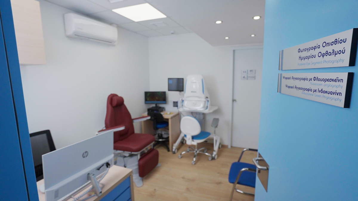
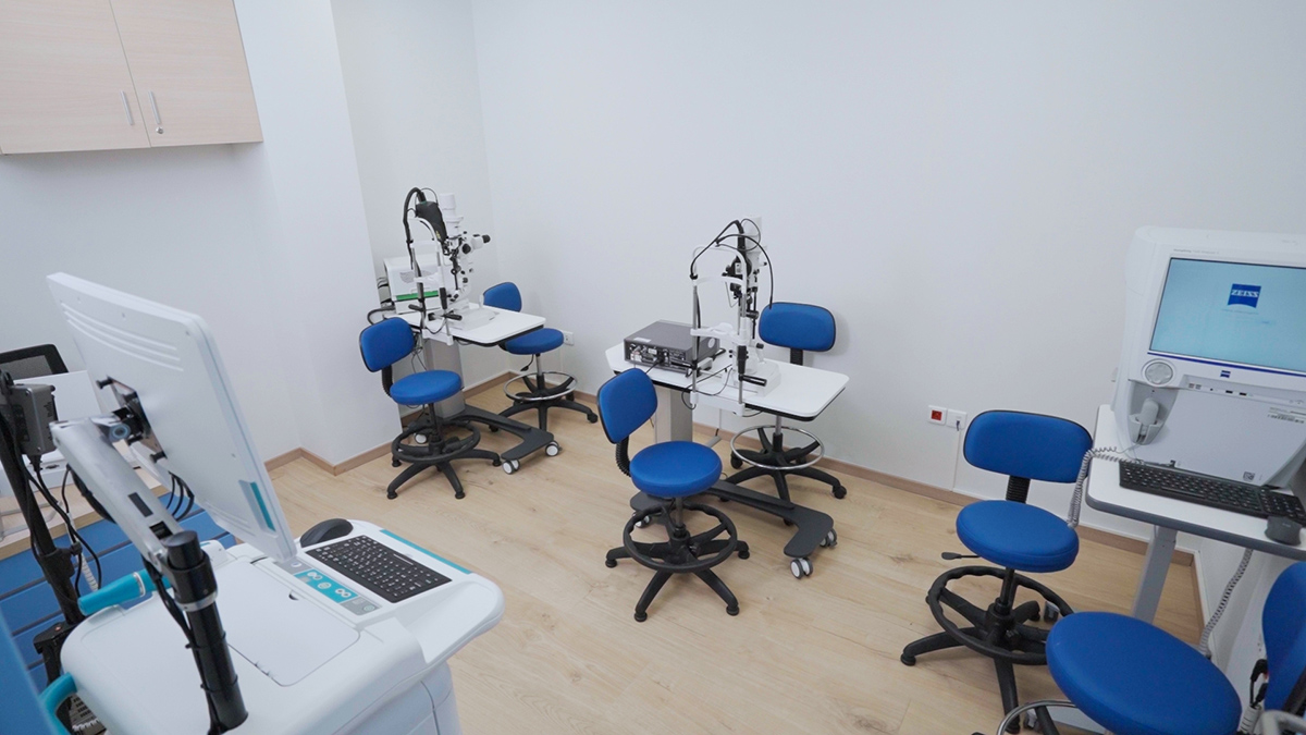
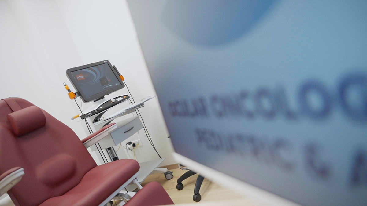
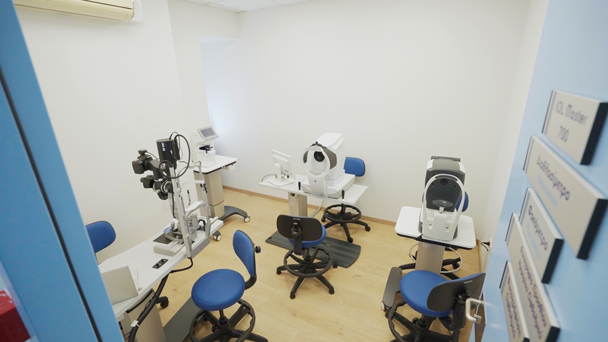
The Ocular Oncology Center for Children & Adults of MITERA is one of the leading ophthalmic oncology units in Europe. We have state-of-the-art technology, ensuring accurate diagnosis and effective treatment.
Under the direction of Dr. Maria Pefkianaki, our specially trained scientific team undertakes the evaluation, diagnosis and treatment of benign and malignant ocular tumors in adults and children.
Dr. Pefkianaki, Director of the Ocular Oncology Center, was trained at the world-renowned Ocular Center, the largest ophthalmic oncology clinic in the world at Wills Eye Hospital, with Dr. Carol Shields and professors who are world-renowned pioneers in the diagnosis and treatment of pediatric and adult patients with ocular tumors.
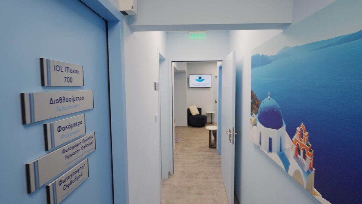
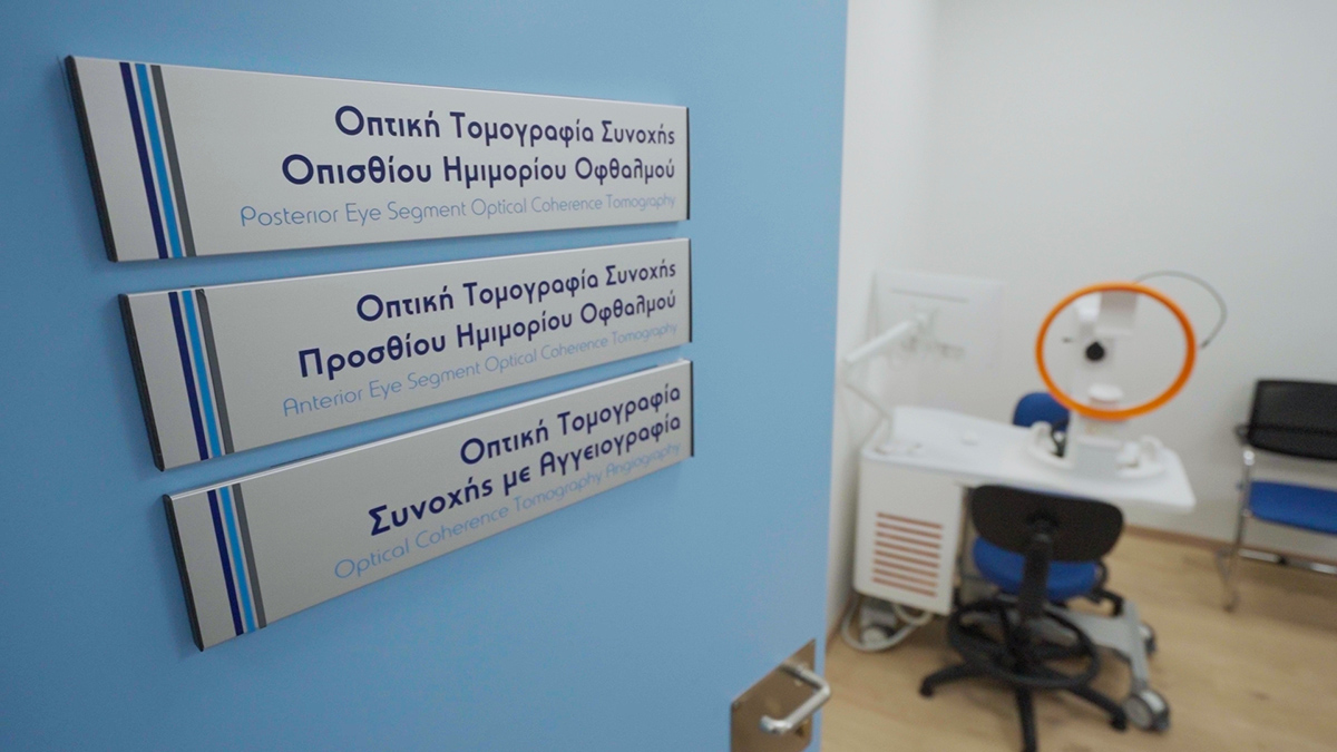
Dr. Pefkianaki and her team are committed to providing a specialized environment that combines scientific training with state-of-the-art technology. We have high-resolution and high-definition ophthalmic equipment, many of which are unique in Greece.
Dr. Pefkianaki, a medical specialist in Ocular Oncology, has established strong partnerships with the world’s leading ocular oncology centers. This gives our patients the opportunity to participate in new research programs and receive the most advanced treatments.
We work closely with physicians of various specialties, such as Oncologists, Radiotherapists, Neuroradiologists, Pediatric Ophthalmologists, Oculoplastic Surgeons, Ophthalmic Geneticists and Social Workers, to provide comprehensive care.
At our center, ocular oncology consultations are held for children and adults with ocular tumors, with the participation of doctors of various specialties. In these consultations, the data are analyzed and the best possible individualized treatment is decided for each patient.
Families with children suffering from retinoblastoma now have access to all aspects of treatment in one location, close to home, without the need to travel abroad.
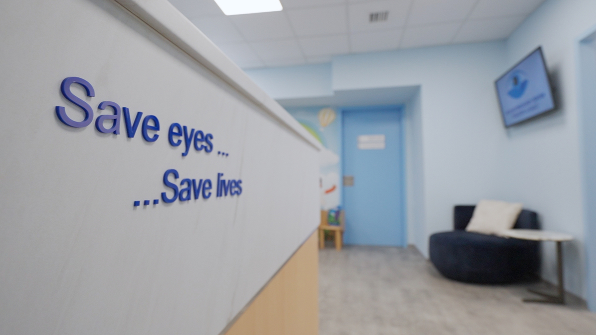
Our mission is to preserve the vision and lives of adult and pediatric patients with ocular tumors.
What to bring to your appointment
In the case of an adult patient with ocular oncology conditions
After checking-in with our receptionist, your visit will continue with the following steps:
In the case of a pediatric patient with ocular oncology conditions
Depending on the age of the child, it is possible that during the visit an ophthalmological examination under general anesthesia may need to be scheduled to thoroughly investigate and exclude or confirm certain ocular diseases.
During the examination under general anesthesia, it is also possible that the following diagnostic tests may need to be performed:
In ocular oncology patients, laser retinal photocoagulation can be used in the case of radiation-induced retinopathy, to destroy or limit retinal ischemia caused by radiation in the eye, or to treat radiation-induced maculopathy.
We provide genetic counseling and testing for prevention and treatment with gene analysis to better assess risk of ocular tumors, particularly retinoblastoma and choroidal melanoma.
Personalized treatments with a patient-centered approach
The patient-centered Ocular Oncology Center for Children & Adults has created Protocols that help patients understand their condition and its treatment.
These include:
** Our center provides separate clinical days for Pediatric and Adult Ocular Oncology Patients.
In addition, there are separate different Operating Rooms for Adult and Pediatric Oncology cases.
Centers of Excellence